Wednesday, June 30, 2010

Rang and Dale just love them some asthma

Heya, short post today as I've been having a social life and all that jazz you do on holidays. This is the respiratory section of Rang & Dale. And by respiratory I really just mean asthma.

So the first 80% of Rang & Dale is just the pathogenesis of Asthma. In short, when susceptible people are exposed to allergens, CD4 gets upset, it produces Th0, which produces Th2, which attracts granulocytes which inflame the bronchi.
Bronchodilators: B2 receptor agonists, muscularinic receptor antagonists, xanthines and cysteinyl leukotriene receptor antagonists. All are smooth muscle relaxants.
Even Rang and Dale couldn't be bothered explaining the immunosuppressant mechanisms of glucocorticoids. Just know that they quash basically all immune response and cause Cushings Syndrome.
Rang and Dale saw fit to cover a brief section on other resp conditions. Treat allergic emergencies with adrenaline, COPDh as little pharmaco management and is largely irreversible, surfactant is important in babies breathing and cough medications are largely useless.

Told you it was short, I don't even have a humourous unrelated drawing to mitigate it. So instead here is a picture of a cat with a top hat and bowtie.
I hope this is in some way pleasing to you.

Tuesday, June 29, 2010

FYI Heparin doesn't really go in your face

This is where it all started, I was locked outside Friend's house waiting for her to return for our group study session for which I was the only one remotely on time. I decided I'd start studying anyway, despite the fact it was cold like the grave. As you've probably quickly noticed, I enjoy playing with words and anti-thrombin III just rearranged itself in my head into Aunty From Bin III, and by the time Friend arrived and let me in I was madly scribbling pictures as fast as I could.
Anti-thrombin III is the main agent against clotting.
Heparin interacts with Anti-thrombin III and inhibits factors 9-12a and thrombin II
Thrombin II cleaves fibrinogen into fibrins and also aggregates platelets, stimulates cell proliferation and modulates smooth muscle contraction
Vitamin K makes factors 2,7,9 and 10. The common F2A channels in Australia.
Heparin -IV, fast, short term. Warfarin - Oral, slower, longer term
Heparin can cause haemorrhage, thrombosis (oddly), hypoaldosteronism and osteoporosis
Heparin IV onset is almost immediate, with s/c it takes about an hour, and it takes 40-90 minutes to wear off. Low Molecular Weight heparin takes longer.
Warfarin is a Vitamin K antagonist
Warfarin is absorbed by the gut, and therefore administered orally, is teratogenic, can occasionally be hepatotoxic, can cause haemorrhage and have a narrow therapeutic window.
I don't really understand this, but those are the basic principles.

Asprin inhibits production of TXA2 which promotes aggregation and promotes production of PCI2 which inhibits platelet aggregation.
Dipyridamole is a phosphodiesterase inhibitor, but just remember it's an anti-platelet drug.
Thienopyridine derivatives (Ticlopidine and clopidogrel) inhibit ADP-dependent aggregation
Seriously. Abciximab? Who the hell came up with that name? Were they trying to summon Cthulhu?
But ridiculous name aside, abciximab, along with tirofiban and eptifibatide, are often given as adjuncts to heparin and aspirin, to reduce restenosis. All of these are glycoprotein IIB/IIIA receptor antagonists.
The fibrinolytic system is activated at the same time as the coagulation system, and plasminogen is deposited on fibrin strands.
Streptinokinase, alteplase, duteplase and reteplase are fibrinolytic drugs with slightly different mechanisms, but in practise they have all been shown to have equal long term outcomes.

Monday, June 28, 2010

It's a total eclipse of the voltage gated calcium channel

This is a short one, most the the drugs that you would think act on the heart are covered under other categories.
Anti-Arrhythmics are broken down into four classes. Class I drugs act by blocking sodium channels, and are divided into three sub classes. Ia (Quinidine, Disopyramide and Procainamide), Ib (Lignocaine (also an anaesthetic) and Phenytoin) and Ic (Flecainide and Encainide). Ib drugs associate and disassociate rapidly, Ic drugs more slowly, and Ia have properties in between. Class II are the beta blockers, and they all end in -olol. Amidarone and Sotalol are Class III and they extend action potential of the heart through unclear mechanisms that somehow affect K channels. Finally Verapamil and Diltiazem are Class IV which block calcium channels and slow conduction of the SA and AV nodes.
Digoxin is a cardiac glycoside derived from foxglove (digitalis), it blocks Na/K channels, which slows down AV conduction. This causes the heart to fill more with each contraction, causing it to contract harder and resulting in a greater ejection fraction. It is used in heart failure and as an antiarrythmic, but it is not a particularly pleasant drug.Nitrates are the most common anti anginal drug. They're a smooth muscle relaxant which allows greater cardiac perfusion. They do however have side effects such a postural hypertension and development of tolerance.
And finally calcium channel blockers (antagonists) as oral antihypertensives. These are grouped under the heart because they have a greater effect on the heart that other smooth muscle relaxants do. As described before they are Class IV anti-arrhythmics, but they also cause the arteries to dilate, with little effect on the veins.

That's it for heart pharmacology, for those of you wondering how the magic of Manic Grandiosity happens below is a portrait of my sister and I 'working together' to construct this blog.

Scanners are the bane of my existence

I was trying to get this done last night, but I ended up being ridiculously late and I gave up. Why was I up so late? Is it because I stayed up to watch the soccer? Nope. Is it because I wasted ages on Facebook? Well, yes, a little bit. But mainly it's because scanners hate me. Like it's not annoying enough having to lift the lid remove the old sheet, place new one in, adjust its position, close the lid, tell the computer to start scanning, and then have a concentration span long enough not to get distracted while the page scans, scanners seem to enjoy being 'helpful' and cropping the pictures down to what it deems as the necessary sections of the picture. It is always wrong in its judgement.
Of course drawing this picture made it take longer and I had to scan it, but I don't pretend for a second to be a rational person.

Anyway this is a little different to what I planned on doing (apparently all my posts come with disclaimers about how not to expect more posts like them :P) this is how I explained the kidney to my friends, but in that case the diagrams were only an aid, rather than the focus, and most of it was me talking. What this verbose vichyssoise ironically attempts to convey is that reducing verbiage is the aim of this project and so I have attempted to remove as much of the associated chatter as possible. We'll start with the gross anatomy.
Put simply, the kidney is divided into a number of lobes, each with a renal pyramid (the medulla) and its associated cortex. Each of the renal pyramids (the peak of which is called a papilla) drains into a minor calyx (calyx meaning cup) which drains into a major calyx, which drains into the renal pelvis and drains into the ureter.
Seriously, the circulation of the kidney makes sense. It's a nice change. The artery that goes to the kidney is called the renal artery. It divides into segments called segmental arteries. Between each lobe there is an interlobar artery. These split at a 90 degree arc into the arcuate arteries. They then shoot off interlobular arteries. That's the only bit that's slightly confusing. And the veins all have the same names. Isn't it nice when the people naming things are being logical instead of artistic or egotistic?
The functional unit of the kidney is the nephron, that's what it looks like . Note that the afferent arteriole is bigger than the efferent arteriole, it's important for maintaining a pressure gradient.
The high hydrostatic pressure of the blood in the glomerulus pushes the blood out into the glomerular capsule. It has to be filtered through the fenestrations in the leaky blood vessels of the glomerulus, which stops large particles including red blood cells. The basement membrane is a gel that acts like a negatively charged sponge, small particles (like water) come through it fine, but large particles (like sand in our sponge metaphor) are filtered, though a few may make it through, it's negativity also keeps out small negatively charged particles like albumin. The final barrier is the podocytes, whose legs wrap around glomeruli and interdigitations (laced fingers) form slits that keep out large particles.

The regulation of filtration rate is quite important. And there are three main regulatory methods. The first is the myogenic reflex, when the arterioles are stretched by a high blood pressure they respond by constricting, which lessens the blood flow into the glomerulus keeping the filtration rate steady. The second is tubular feedback, the mechanisms of which aren't entirely clear, but when the PCT detects a lower or higher flow the juxtaglomerular cells constrict or relax the arterioles to change the blood flow. The final method is the renin-angiotensin cycle, the kidney secretes renin, which converts angiotensin to angiotensin I, which is converted by ACE into angiotensin II. Which acts on the brain, vessels, adrenal glands and kidneys, stimulating thirst, raising blood pressure secreting aldosterone and increasing NaCl and H2O retention respectively.
Proximal Convoluted Tubular re-absorption is driven by sodium. The cells which line the tubule constantly pump out Na+ keeping the concentration low, causing Na+ to diffuse across the membrane. Glucose is bound to Na+ by transport proteins, causing it to follow along, along with negatively charged molecules such as chlorine . As the osmolarity changes water also diffuses out. Bicarbonate is unable to be absorbed, but none is usually found in the urine, so within the cell water and CO2 combine to form bicarbonate and H+. The H+ moves into the tube and breaks the bicarbonate into water and CO2, which can diffuse out.

This is possibly one of the coolest things ever. No really, I know it's nerdy. But seriously, the thin part of the nephron loop is permeable only to water, so when it dips into the salty medulla the water leaves the tube and the osmolarity increases. When it reaches the thick part which is non-permeable to water the concentration of sodium has increased, and so as it leaves the medulla the sodium bails as well, preserving the sodium gradient.
Now think about this, what would happen if the vessels went straight through the medulla? The salt would go in and the water would go out. Very very bad. Hence the vasa recta, they go enter and leave the medulla next to each other and thus as the gradient increases the salt enters and the water leaves, but as the vasa recta returns more or less on its original path the reverse occurs, meaning the osmolarity and concentration is essentially unchanged.
Finally we reach the Distal Collecting Tubule and Collecting Duct, these remove the last of the water and the salt, and are modulated by aldosterone, anti-natriuretic peptide, antidiuretic hormone and parathyroid hormone depending on whether or not the body needs to conserve or excrete water. The method of the collecting duct is similar to the thin section of the nephron loop, as the medulla becomes saltier more water leaves the duct.

And that's the kidney! It actually makes a lot of sense and it's a really cool organ.

Friday, June 25, 2010

The Cranial Nerves

The cranial nerves are one of those things that don't agree with me, because they're essentially a list of forms and functions. We could try to remember them like this...

I - Olfactory Nerve - Supplies sensory fibres to the nose, to the olfactory mucosa in the upper parts of the nasal cavity, instrumental in sense of smell, runs through the cribriform plate.

But the olfactory nerve is one of the simplest cranial nerves and trying to learn twelve just like it would kill me. So I've drawn a series of pun heavy pictures instead. Not all my posts will be this picture-pun heavy, but anatomy is a tricky subject to represent comically.


Olfactory Nerve - Sense of Smell - Cribriform Plate


Optic Nerve - Information from eye to the brain - Optic Canal


This one needs to be redone, the Occulomotor nerve runs through the Tentorium but it doesn't go through the optic canal, it runs through the Superior Orbital Fissure. It then supplies all the muscles that move the eye except the Lateral Rectus and Superior Oblique. It is responsible for the pupillary reflex.


Trochlear Nerve - Superior Orbital Fissure - Superior Oblique


Trigeminal Nerve is where thing start to get a little complicated. It includes three parts, the Opthalmic (runs through Superior Orbital Fissure), the Maxillary (Runs through Foramen Rotundum) and the Mandibular (Runs through Foramen Ovale). Only the mandibular has a motor supply (It supplies the muscles of mastication as well as a few others involved in chewing biting and swallowing), the others are purely sensory.


Abducens - Superior Orbital Fissure - Lateral Rectus


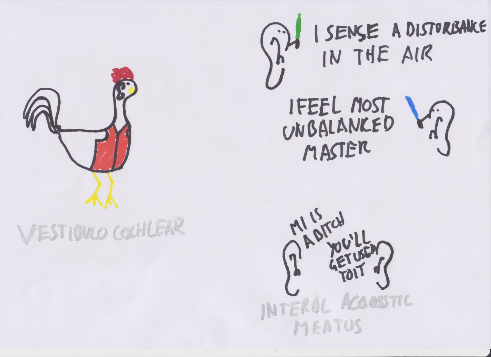
The Facial Nerve supplies the muscles of facial expression, the corneal reflex and (via the chorda timpani) taste in the anterior two thirds of the tongue and stimulation of the submandibular and sublingual salivary glands. It runs through the Internal Auditory Meatus.


Vestibulocochlear - Sound, balance and orientation - Internal Acoustic Meatus (What the ears are saying is irrelevant, I just didn't want to repeat the joke.)


It really doesn't sound like anything punny, I swear. I tried and tried. Supplies taste to the anterior two thirds of the tongue and the parotid salivary gland. Runs through the Jugular Foramen.


Vagus Nerve - Supplies muscles of the pharynx and larynx - Jugular Foramen


Accessory nerve - Supplies Trapezius and Sternocleidomastoid (which allowing shrugging and head movement - Jugular Foramen


Hypoglossal Nerve - Supplies the muscles that move the tongue - Hypoglossal Canal


Being able to know which of the nerves have a motor or sensory supply can be helpful to remember the specifics of what they do. The above mnemonic tells you whether they have Motor (M), Sensory (S) or Both (B).


I don't like being this blatantly vulgar, I like to be a little more subtle about it, but the most popular mnemonics are dirty ones, and there wasn't a PG equivalent. I'm also not hugely keen on acronym mnemonics, but the holes the nerves go through are a pain to remember, so I found a backup to just my pictures to be helpful. Translation is as follows

Cribriform Plate I, Optic Canal II, Superior Orbital Fissure III, Superior Orbital Fissure IV, Superior Orbital Fissure V1, Foramen Rotundem V2, Foramen Ovale V3, Superior Orbital Fissure VI, Internal Acoustic Meatus VII, Internal Acoustic Meatus VIII, Jugular Foramen IX, Jugular Foramen X, Jugular Foramen XI, Hypoglossal Canal XII

That's it for the cranial nerves, I hope it's at least some help to you. The next one I'll draw and upload will probably be my kidney tutorial derived from Saladin. I have working sets of notes for Rang and Dale for Chapters 18, 21, 23 & 24 but I want to do a bit more work on the before I upload them.

Edit: If anyone has any specific requests for topics they'd like me to cover just comment/email/whatever me and I'll prioritise that.